Skip Navigation
Skip to contents

JYMS : Journal of Yeungnam Medical Science

Indexed in: ESCI, Scopus, PubMed,
PubMed Central, CAS, DOAJ, KCI
FREE article processing charge
OPEN ACCESS
SEARCH
Search

Articles

Page Path
HOME > J Yeungnam Med Sci > Volume 40(Suppl); 2023 > Article
Resident fellow section: Teaching images
Ultrasound-guided diagnosis/intervention for ischiofemoral impingement syndrome
Wei-Ting Wu1,2orcid, Ke-Vin Chang1,2,3orcid, Levent Özçakar4orcid
Journal of Yeungnam Medical Science 2023;40(Suppl):S134-S136.
DOI: https://doi.org/10.12701/jyms.2023.00500
Published online: July 12, 2023

1Department of Physical Medicine and Rehabilitation, National Taiwan University Hospital, Bei-Hu Branch, Taipei, Taiwan

2Department of Physical Medicine and Rehabilitation, National Taiwan University College of Medicine, Taipei, Taiwan

3Center for Regional Anesthesia and Pain Medicine, Wang-Fang Hospital, Taipei Medical University, Taipei, Taiwan

4Department of Physical and Rehabilitation Medicine, Hacettepe University Medical School, Ankara, Turkey

Corresponding author: Ke-Vin Chang, MD, PhD Department of Physical Medicine and Rehabilitation, National Taiwan University Hospital, Bei-Hu Branch, No. 87, Nei-Jiang Rd., Wan-Hwa District, Taipei 108, Taiwan Tel: +886-2-23717101-5309 • E-mail: kvchang011@gmail.com
• Received: May 18, 2023   • Revised: May 27, 2023   • Accepted: June 2, 2023

Copyright © 2023 Yeungnam University College of Medicine, Yeungnam University Institute of Medical Science

This is an Open Access article distributed under the terms of the Creative Commons Attribution Non-Commercial License (http://creativecommons.org/licenses/by-nc/4.0/) which permits unrestricted non-commercial use, distribution, and reproduction in any medium, provided the original work is properly cited.

  • 949 Views
  • 74 Download
A 54-year-old man complained of right buttock pain that had persisted for 2 weeks and worsened when walking at a fast pace or climbing stairs. He did not experience pain radiating to the ipsilateral leg and denied any sports injury or trauma to his buttocks. Treatment with nonsteroidal anti-inflammatory drugs and physical modalities (e.g., ultrasound diathermy and transcutaneous electrical nerve stimulation) did not alleviate his symptoms. With a suspected diagnosis of piriformis syndrome, the patient was referred to the Department of Physical Medicine and Rehabilitation for an ultrasound-guided injection.
During ultrasound examination of the right gluteal region, the piriformis muscle and tendon were found to have normal echotexture and size. Sonopalpation did not elicit pain, and the patient reported that the pain seemed to originate in a region caudal to the great sciatic notch. The ultrasound transducer was then moved toward the ischial tuberosity. Compared to the asymptomatic side (Fig. 1A), swelling and loss of the fibrillary pattern over the medial aspect of the quadratus femoris muscle were detected on the symptomatic side (Fig. 1B). Power Doppler imaging revealed a normal medial femoral circumflex artery between the quadratus femoris and obturator externus (OE) muscles without increased intramuscular vascularity (Fig. 2A). Because the patient experienced buttock pain during internal/external hip rotation, ischiofemoral impingement syndrome (IFS) was highly likely the underlying cause. Using an in-plane approach, ultrasound-guided injection of the affected quadratus femoris muscle was performed using a mixture of 5 mL 50% dextrose and 5 mL 1% lidocaine (Fig. 2B). Two weeks after the injection, the patient reported complete pain relief.
IFS results from narrowing of the space between the ischial tuberosity and trochanter, leading to irritation of the quadratus femoris muscle [1]. Patients typically present with posterior buttock pain and the clinical scenario cannot be distinguished from that of deep gluteal and hamstring syndromes. IFS is associated with several biomechanical factors including femoral anteversion with a compensatory toe-in posture during ambulation [2]. Physical findings may include tenderness near the ischial tuberosity and a positive long-stride walking test [3]. Magnetic resonance imaging (MRI) is often necessary to confirm the diagnosis, as it can reveal edema, fatty infiltration, and increased thickness of the quadratus femoris muscle [4].
Ultrasonography can be useful for visualizing deep gluteal muscles, including the quadratus femoris [5]. However, because a large portion of the quadratus femoris muscle is hidden by the acoustic shadow of the ischial tuberosity, not every patient with IFS exhibits pathological sonographic findings. However, internal femoral rotation improves visualization of the muscle's medial aspect [6]. Ultrasound-guided injection of the fascial plane between the quadratus femoris and the OE muscles can facilitate the diagnosis and treatment of IFS by allowing more injectate to infiltrate the deep portion of the quadratus femoris. It is important that power Doppler imaging is used during injection to prevent collateral injury to the medial femoral circumflex artery that typically courses inside the target fascial space. In cases where patients do not respond to ultrasound-guided interventions or experience recurrent pain shortly after injection, it is advisable to promptly schedule an MRI to exclude the possibility of a more serious condition such as sarcoma of the pelvic cavity.
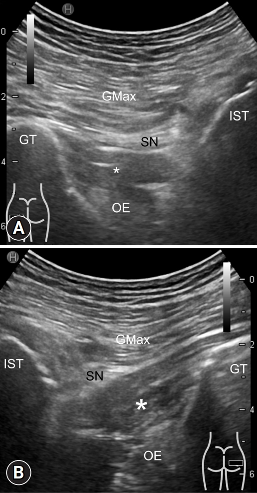
• Ischiofemoral impingement syndrome is caused by narrowing of the space between the ischial tuberosity and trochanter, leading to posterior buttock pain.
• Magnetic resonance imaging is the gold standard for diagnosis, whereas ultrasonography can help visualize the deep gluteal muscles.
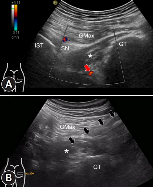
• Ultrasound-guided injection between the quadratus femoris and obturator internus is useful in treatment but requires power Doppler imaging to prevent collateral vascular injury.

Ethical statements

In the authors’ institutions, approval from the institutional review board is not required for the case report. Written patient consent was obtained for the publication of this report.

Conflicts of interest

Wei-Ting Wu and Ke-Vin Chang have been editorial board members of the Journal of Yeungnam Medical Science since 2021. They were not involved in the review process of this manuscript. There are no other conflicts of interest to declare.

Funding

This work was funded by the National Taiwan University Hospital, Bei-Hu Branch; Ministry of Science and Technology (MOST 106-2314-B-002-180-MY3 and 109-2314-B-002-114-MY3); and the Taiwan Society of Ultrasound in Medicine.

Author contributions

Conceptualization: all authors; Investigation, Data curation: WTW, KVC; Formal analysis, Funding acquisition, Supervision: KVC, LÖ; Validation: LÖ; Visualization: KVC; Writing-original draft: KVC; Writing-review & editing: all authors.

Fig. 1.
Ultrasound findings of the left and right gluteal regions. In contrast to (A) the left healthy side, (B) enlargement and absence of the fibrillary pattern in the medial region of the right quadratus femoris muscle (asterisk) is seen. GMax, gluteus maximus; IST, ischial tuberosity; SN, sciatic nerve; GT, greater trochanter; OE, obturator externus.
jyms-2023-00500f1.jpg
Fig. 2.
(A) Power Doppler ultrasound image shows the right medial femoral circumflex artery (red arrow) seen between the quadratus femoris (asterisk) and obturator externus muscles. (B) Using an in-plane approach, ultrasound-guided injection of 5-mL 50% dextrose and 5-mL 1% lidocaine was administered into the right quadratus femoris muscle (asterisk). Arrows, needle trajectory. GMax, gluteus maximus; IST, ischial tuberosity; SN, sciatic nerve; GT, greater trochanter.
jyms-2023-00500f2.jpg
  • 1. Wu WT, Chang KV, Mezian K, Naňka O, Ricci V, Chang HC, et al. Ischiofemoral impingement syndrome: clinical and imaging/guidance issues with special focus on ultrasonography. Diagnostics (Basel) 2022;13:139.ArticlePubMedPMC
  • 2. Scorcelletti M, Reeves ND, Rittweger J, Ireland A. Femoral anteversion: significance and measurement. J Anat 2020;237:811–26.ArticlePubMedPMCPDF
  • 3. Gómez-Hoyos J, Martin RL, Schröder R, Palmer IJ, Martin HD. Accuracy of 2 clinical tests for ischiofemoral impingement in patients with posterior hip pain and endoscopically confirmed diagnosis. Arthroscopy 2016;32:1279–84.ArticlePubMed
  • 4. Akça A, Şafak KY, İliş ED, Taşdemir Z, Baysal T. Ischiofemoral impingement: assessment of MRI findings and their reliability. Acta Ortop Bras 2016;24:318–21.ArticlePubMedPMC
  • 5. Chang KV, Wu WT, Lew HL, Özçakar L. Ultrasound imaging and guided injection for the lateral and posterior hip. Am J Phys Med Rehabil 2018;97:285–91.ArticlePubMed
  • 6. Mezian K, Ricci V, Güvener O, Jačisko J, Novotný T, Kara M, et al. EURO-MUSCULUS/USPRM dynamic ultrasound protocols for (adult) hip. Am J Phys Med Rehabil 2022;101:e162–8.ArticlePubMed

Figure & Data

References

    Citations

    Citations to this article as recorded by  

      Figure
      Related articles

      JYMS : Journal of Yeungnam Medical Science